South Australian Medical Heritage Society Inc
Website for the Virtual Museum
Home
Coming meetings
Past meetings
About the Society
Main Galleries
Medicine
Surgery
Anaesthesia
X-rays
Hospitals,other organisations
Individuals of note
Small Galleries
Ethnic medicine
- Aboriginal
- Chinese
- Mediterran
Histology - History and BasicsACKNOWLEDGMENTS; The photographs and information about the techniques has kindly been provided by Linda Wurfel and Dr. Lynette Moore (head) of the Department of Histology at the Women and Children’s Hospital Adelaide. Some of the items and samples date back to the early 1900s.
The OED mentions “histo” a Greek word meaning web or tissue. In combination with an identifying word the meaning changes. Thus histo “blast” meaning a primitive cell, or histo “logy” indicating the study of the minute structure of animal or plant tissues usually using a microscope. The OED attributes the word to Craig in 1847.
In medical practice Histology is invaluable in confirming the clinical diagnosis. It is now unlikely that treatment of a malignancy or other diseases is undertaken without a biopsy and microscopy.
In order to prepare tissue for such a process a small sample is taken from the abnormal area, placed (“fixed”) in 10% formaldehyde and sent to the laboratory for processing.

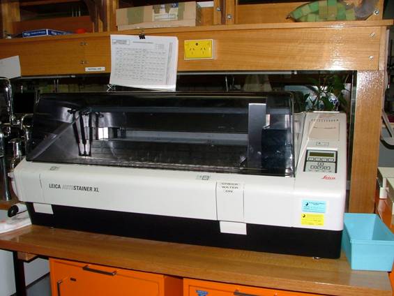
An automatic processing machine with sequential concentrations of alcohol and xylene
In order to dehydrate the tissue the sample is placed in increasing concentrations of Ethanol and then cleared with Xylene. Finally it is is impregnated with paraffin which replaces the Xylene. The whole process may take 12 hours. When the process is completed the tissue is embedded in a paraffin block.
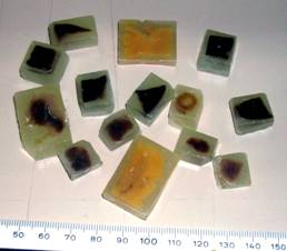
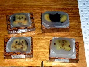
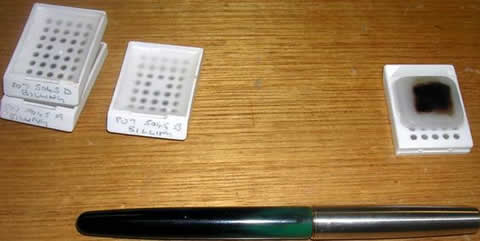
The currently used blocks are smaller and the preparation is partly automated. They are commonly called cassettes.
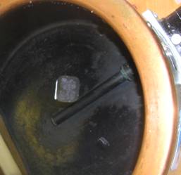
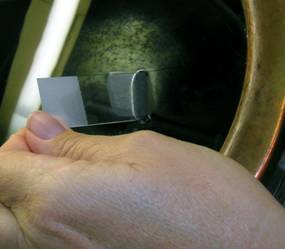
To be able to examine the tissue embedded in paraffin under a microscope it has to be very thin and the cells need to be stained with special dyes in order to identify the nuclei and other cell components. The tissue sections are cut with a microtome. The section thickness usually ranges between 2-5 microns (2-5 thousandths of a millimetre).
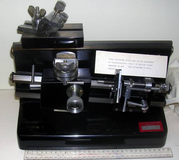
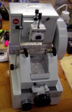
An early microtome
used 50 years ago (1954)
A contemporary microtome
.
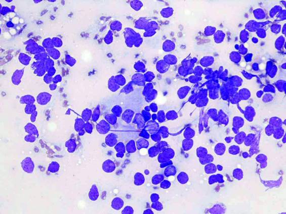
A very popular stain is Haematoxylin and Eosin. The former stains the cell nuclei blue while Eosin stains the cytoplasm pink. There are numerous other more specific stains, which can selectively stain specific cellular components. Many originated from textile dyes.
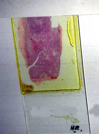
A haematoxylin eosin slide
In order to see the cellular structure of the tissue on the slide a microscope is required The first indication of enlarging a structure may well have been an accidental finding of a biconvex piece of glass. The magnification may have been no more than x4 but using more lenses in sequence produced surprising results. Anton van Leeuwenhoek (1632-1723) is thought to be one of the early inventors of the microscope. He used a combination of lenses and at this early time was able to achieve a magnification of 270x.Thus it was possible to identify individual cells and show the structure of different tissues.

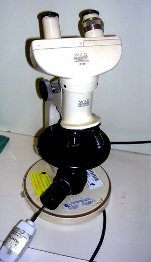
Early microscope (from Google) Modern binocular microscope
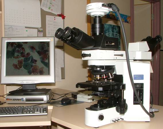
Present day binocular microscope with a digital camera & computer
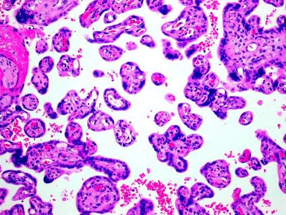
Haematoxylin & eosin stain showing placenta
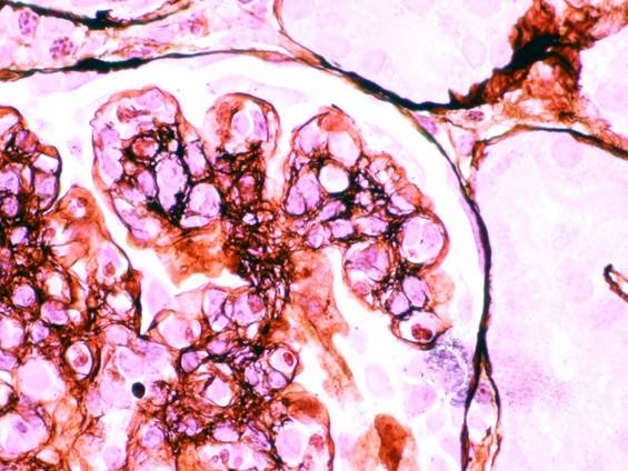
Kidney biopsy silver stain
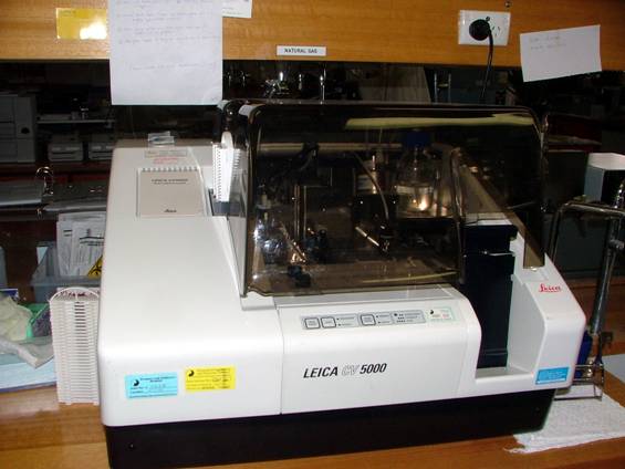
More recently automation has produced machines
which stain slides and apply cover slips
Modern automated slide stainer
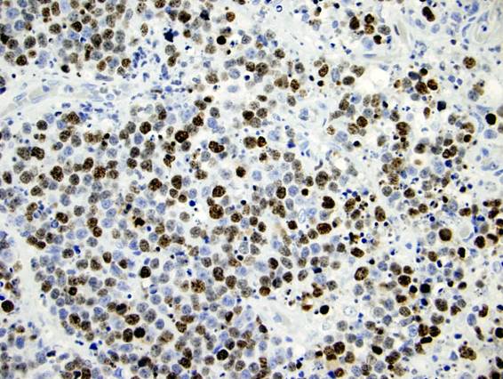
Immuno-peroxdise stain
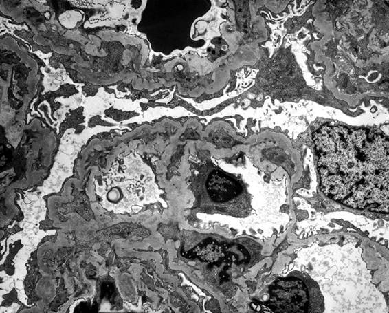
Electron microscopy of renal biopsy
-o0o-